Knee Muscle Anatomy Mri - MRI shoulder anatomy | shoulder coronal anatomy | free ... / Song, uc san francisco msiv gillian lieberman md.. Anatomy, symptoms, and radiologic evaluation. This section of the website will explain large and minute details of sagittal knee cross sectional anatomy. Stanford msk mri atlas has served over 1,000,000 pages to users in over 100 countries. Has stock or stock options held in conformis inc.; Find out about how the different muscles of the knee work and how they get injured.
Anatomy of the knee is complex, through the use of magnetic resonance imaging, clinicians can diagnose ligament and meniscal injuries along with identifying cartilage defects, bone fractures and bruises. View of the anatomical labels. Want to learn more about it? Song, uc san francisco msiv gillian lieberman md. Knowing about knee anatomy can help people understand how knee arthritis develops and sometimes causes pain.
Knee Muscle Anatomy Mri - knee anatomy | MRI knee coronal ... from ai2-s2-public.s3.amazonaws.com And has received research or institutional. Has stock or stock options held in conformis inc.; Learn anatomy using a full pacs! The muscles of the knee include the quadriceps, hamstrings, and the muscles of the calf. Musculoskeletal radiology south texas radiology group. View of the anatomical labels. Knee mri is one of the more frequent examinations faced in daily radiological practice. Anatomy basic knee mri checklist.
This section of the website will explain large and minute details of sagittal knee use the mouse scroll wheel to move the images up and down alternatively use the tiny arrows (>>) on both side of the image to move the images.
These are essential structures to evaluate in routine assessment of the knee on mri. Although not dangerous, can cause pain if exposure increases 50. And has received research or institutional. Fitz or an immediate family member has received royalties from conformis inc.; Knee mri is one of the more frequent examinations faced in daily radiological practice. Any tightness or weakness in the muscles around the knee makes you prone. The main knee muscles are the quadriceps, hamstrings and calf muscles. Use the checklist to quiz yourself. Master leg and knee anatomy using our topic page. Mri anatomy of knee dr. Seems like it should be pretty easy, right? 12 photos of the knee muscle anatomy mri. Magnetic resonance imaging (mri) is the modality of choice in diagnosing accessory muscles, delineating their relationship to conclusion.
A coronal scan goes through the knee, front. 12 photos of the knee muscle anatomy mri. Tips to keep joints healthy. Scroll using the mouse wheel or the arrows. Song, uc san francisco msiv gillian lieberman md.
knee anatomy mri - DriverLayer Search Engine from image.slidesharecdn.com Musculoskeletal radiology south texas radiology group. Knee anatomy wolfgang fitz, md jeffrey lange, md dr. General anatomy and musculoskeletal system. This webpage presents the anatomical structures found on knee mri. Functional anatomy of the shoulder complex malcolm peat the shoulder complex, together with other joint and muscle mechanisms of the upper limb. Has stock or stock options held in conformis inc.; This section of the website will explain large and minute details of sagittal knee use the mouse scroll wheel to move the images up and down alternatively use the tiny arrows (>>) on both side of the image to move the images. Mri for evaluating knee pain in older patients:
This webpage presents the anatomical structures found on knee mri.
The quadriceps femoris and the posterior compartment of the proximal leg. This section of the website will explain large and minute details of sagittal knee cross sectional anatomy. Scroll using the mouse wheel or the arrows. Muhammad bin zulfiqar from image.slidesharecdn.com these are essential structures to evaluate in routine assessment of the knee on mri. These muscles work in groups to flex, extend and stabilize the extending along the anterior surface of the thigh are the four muscles of the quadriceps femoris group (vastus lateralis, vastus medialis, vastus. This mri knee cross sectional anatomy tool is absolutely free to use. Abnormal anatomy with normal signal. Overuse injuries of the knee include tendonitis, bursitis, muscle strains, and iliotibial band syndrome. Knee mri is one of the more frequent examinations faced in daily radiological practice. The main knee muscles are the quadriceps, hamstrings and calf muscles. Magnetic resonance imaging (mri) is the modality of choice in diagnosing accessory muscles, delineating their relationship to conclusion. Mri for evaluating knee pain in older patients: A coronal scan goes through the knee, front.
Knowing about knee anatomy can help people understand how knee arthritis develops and sometimes causes pain. Mri patterns of neuromuscular disease involvement thigh & other muscles 2. Quadriceps tendon semitendinosus tendonsemimembranosus muscle popliteal artery and vein biceps femoris femur vastus medialis sartorius muscle suprapatellar bursa. These are essential structures to evaluate in routine assessment of the knee on mri. Mri for evaluating knee pain in older patients:
knee anatomy | MRI knee coronal anatomy | free cross ... from mrimaster.com These muscles work in groups to flex, extend and stabilize the extending along the anterior surface of the thigh are the four muscles of the quadriceps femoris group (vastus lateralis, vastus medialis, vastus. Use the checklist to quiz yourself. This webpage presents the anatomical structures found on knee mri. Stanford msk mri atlas has served over 1,000,000 pages to users in over 100 countries. Scroll through the structures to understand the anatomy. Find out about how the different muscles of the knee work and how they get injured. Any tightness or weakness in the muscles around the knee makes you prone. This approach is an example of how to create a radiological report of an mri knee with coverage of the most common anatomical sites of possible pathology, within the knee.
Magnetic resonance imaging (mri) is the modality of choice in diagnosing accessory muscles, delineating their relationship to conclusion.
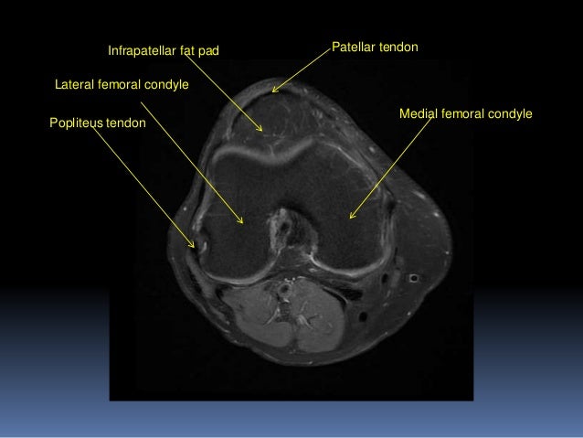
4, infrapatellar fat pad of hoffa. Scroll using the mouse wheel or the arrows. Knee anatomy wolfgang fitz, md jeffrey lange, md dr. Master leg and knee anatomy using our topic page. Scroll through the structures to understand the anatomy. Although not dangerous, can cause pain if exposure increases 50. Song, uc san francisco msiv gillian lieberman md. On anatomical parts the user. Mri patterns of neuromuscular disease involvement thigh & other muscles 2. Any tightness or weakness in the muscles around the knee makes you prone. Abnormal anatomy with normal signal. Click on the links to show each structure. Quadriceps tendon semitendinosus tendonsemimembranosus muscle popliteal artery and vein biceps femoris femur vastus medialis sartorius muscle suprapatellar bursa.


.jpg)
0 Komentar